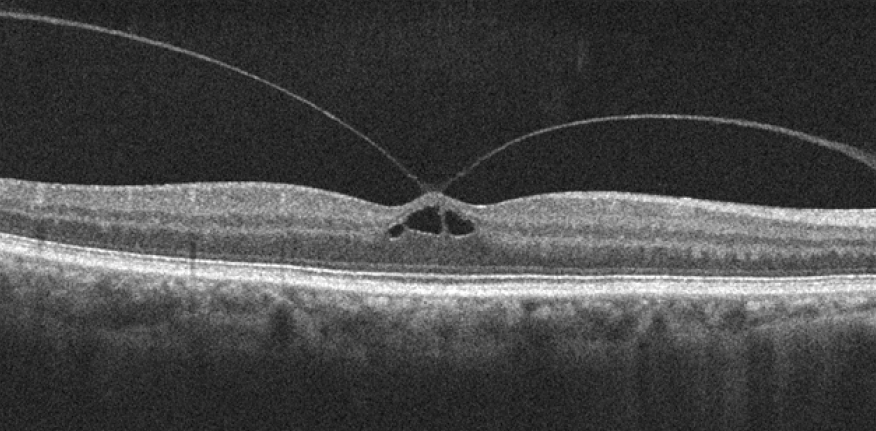
VMT can be classified as focal (≤1500µm) or broad (>1500µm) depending on the diameter of vitreous attachment and as concurrent or isolated based on morphologic finding on OCT images. These glial cells contribute to the contractile forces in VMT. Histopathologic examination of VMT specimens demonstrates a variety of cell types such as astrocytes, myofibroblasts and fibrocytes. The prevalence of isolated idiopathic VMT is approximately 0.6 per 100 000 of the general population. VMT can occur in isolation, or in conjunction with comorbid macular conditions, as macular hole, macular edema, and epiretinal membrane (ERM). Vitreomacular traction (VMT) is defined as posterior vitreomacular attachment with tractional distortion of the perifoveal architecture inducing visual disturbance. Vitreoretinal interface disorders refer to a spectrum of pathologic interactions between the posterior hyaloid and the underlying retinal surface, ranging from innocuous attachment to substantial disruption of retinal integrity. Further studies are needed to evaluate its indications, benefits, and risks. Pneumatic vitreolysis (PVL) with limited face-down position is a viable option for treating focal VMT with few adverse events. One patient developed a retinal break at upper nasal retina after two weeks of injection. The rate of release in phakic eyes was 90% (18 of 20 eyes) versus 60% in pseudophakic eyes (6 of 10 eyes). VMT release was documented on SD-OCT at an average of 3 weeks (range, 1–12 weeks). Overall, VMT release occurred in 24 of 30 eyes by the final follow-up visit (80% final release rate) furthermore, 76.9% of eyes with diabetic maculopathy and 25% of eyes with concurrent epiretinal membrane (ERM) had successful VMT release.

Secondary outcome measures were changes in postoperative BCVA andCFT. Primary outcome measure was release of VMT. Postoperatively, we performed SD-OCT at one week, one month, and three months for all eyes. All eyes received single intravitreal injection of 0.3 mL of 100% SF6 gas. Pre-operatively, mean best corrected visual acuity (BCVA) was 20/125 (range 20/400–20/40).
#VMT RETINA SERIES#
This is a prospective interventional case series including 30 eyes of 29 patients with symptomatic focal VMT evident on SD-OCT.

In Canada, JETREA is indicated for the treatment of symptomatic vitreomacular adhesion (VMA).To evaluate the efficacy of single intravitreal injection of an expansile concentration of sulphur hexafluoride gas (SF6) in treating patients with symptomatic focal vitreomacular traction (VMT) documented by spectral domain optical coherence tomography (SD-OCT) preoperatively. However, ocriplasmin has been developed and is approved by the European Medicines Agency (EMA) for treatment of VMT in adults, including when associated with macular hole of diameter less than or equal to 400 microns under the trade name JETREA®. Until recently, vitrectomy was the only treatment for VMT/VMA and MH.9 Because of the risks associated with vitrectomy (including retinal detachment, hemorrhage, and cataract), vitrectomy is usually only performed once the loss of vision has become clinically significant.Īttempts at developing enzymes for vitreolysis against the substrates responsible for VMA have been generally unsuccessful. VMT can result in macular deformation and macular hole (MH), an opening in the retina that develops at the fovea. This leads to separation of the posterior vitreous cortexįrom the retinal internal limiting membrane – a process called posterior vitreous detachment (PVD). As part of the normal aging process, the protein structure of the vitreous progressively liquefies and the vitreoretinal adhesions weaken.

The consistency of the vitreous and the adhesion to the retina are maintained by a matrix of proteins including collagen, laminin, and fibronectin. Under normal physiologic conditions, the vitreous maintains a gel-like consistency and adheres completely to the entire surface of the retina.


 0 kommentar(er)
